
What Are Cataracts? A Clear Guide to Understanding and Protecting Your Vision
If you are wondering, “What are cataracts?”, the simple answer is that a cataract is the clouding of the eye’s natural crystalline lens. This clouding can make vision seem hazy, dim, glary, or less colourful, and it usually develops gradually over time rather than appearing suddenly.
At The Focal Point Optometrist in Perth, cataract-related vision changes are assessed through a thorough, attentive, and highly personalised approach to care. If you have noticed increasing glare, difficulty with night driving, or spectacles/glasses that no longer feel clear, a comprehensive eye test can help assess your lens clarity, check your broader eye health, and determine whether monitoring or referral to an ophthalmologist may be appropriate.
Key Takeaways:
- Cataracts occur when proteins within the crystalline lens gradually break down and clump together.
- Common symptoms may include painless blur, glare, halos around lights, faded colour vibrancy, and frequent script changes.
- Early cataracts may often be monitored and managed with updated spectacles/glasses, lighting changes, and glare reduction strategies.
- When cataracts significantly interfere with daily life, your optometrist may refer you to an ophthalmologist for surgical assessment.
Cataracts are common with ageing, but they are not the only possible cause of reduced clarity. Regular optometric care can support early detection, monitoring, and timely referral where needed, helping you make informed decisions about your vision and eye health.
What Are Cataracts and How Do They Develop?
A cataract is the progressive clouding of the crystalline lens inside the eye. In a healthy eye, this crystalline lens sits just behind the pupil and iris. Its primary biological function is to flex, adjust focus, and allow light rays to pass cleanly onto the retina at the back of the eye. The retina converts these light signals into nerve impulses, which the brain interprets as crisp, clear images.
The crystalline lens is composed primarily of water and specialised structural proteins called crystallins. In early life, these proteins are arranged in a precise, transparent matrix that allows light to transmit through without obstruction. As the body matures, oxidative stress, cellular wear, and environmental exposures cause these natural proteins to gradually denature, break down, and aggregate together.
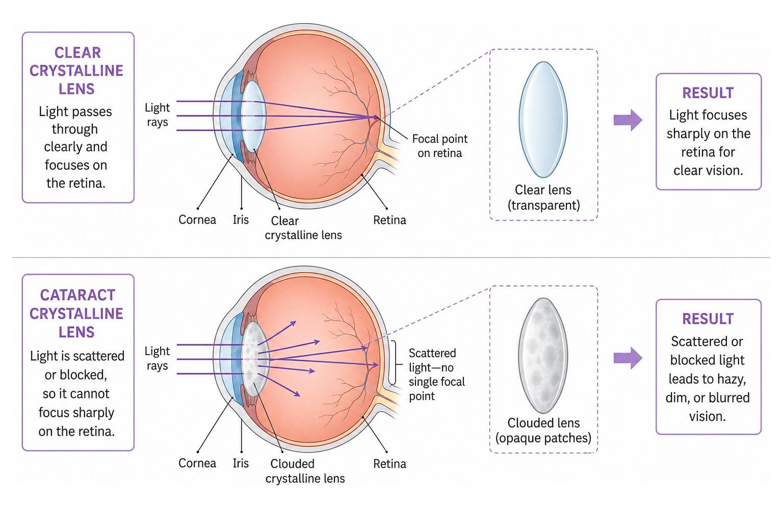
When proteins clump together, they form opaque patches within the lens matrix. Instead of passing smoothly through the clear structure, incoming light rays scatter in multiple directions or become partially blocked. This scattering prevents the retina from receiving a sharp focal point, leading to hazy, washed-out, or dimmed visual clarity.
Protein breakdown in the human lens is a normal part of the physiological ageing process. Microscopic changes within lens proteins can begin as early as adulthood, though early cellular shifts rarely produce immediate symptoms. Over subsequent decades, these protein clusters expand and harden. In older adults, the density of these opacities reaches a point where visual quality gradually begins to decline.
What Factors Increase the Risk of Cataract Formation?
While natural ageing represents the primary underlying factor in cataract formation, several environmental, systemic, and lifestyle influences can accelerate changes within the crystalline lens:
- Ultraviolet (UV) Radiation: Cumulative, unprotected exposure to solar UV radiation generates free radicals within ocular tissues. These reactive molecules accelerate oxidative damage to delicate lens proteins.
- Systemic Health Conditions: Chronic metabolic conditions, particularly diabetes mellitus, can cause elevated glucose levels in the aqueous humor. This excess sugar enters the crystalline lens, causing structural swelling and accelerated protein glycation.
- Ocular Trauma and Surgery: Blunt-force eye injuries, penetrating trauma, or previous intraocular surgical procedures can disrupt the delicate lens capsule, leading to localised or rapid opacification.
- Lifestyle Influences: Tobacco smoking introduces systemic toxins that deplete natural antioxidants in the eye, substantially increasing oxidative stress. High alcohol consumption is also associated with metabolic stress in ocular tissues.
- Medications: Prolonged systemic or topical use of corticosteroid medications can disrupt metabolic transport across the lens capsule, promoting rapid cataract formation.
Adopting proactive visual wellness habits and holistic lifestyle choices can help safeguard delicate ocular structures. For further strategies on supporting ocular vitality, explore our guide to healthy eyes.
What Are the Primary Symptoms and Types of Cataracts?
Because cataracts generally develop slowly, the initial signs can be subtle. Over time, progressive opacification creates recognisable visual disturbances that may affect daily routines:
- Painless Progressive Blurring: A slow, painless loss of visual sharpness at both near and far distances, often described as looking through dusty glass.
- Glare and Light Sensitivity: Heightened sensitivity to bright sunlight, indoor task lighting, or overhead fluorescent globes.
- Night Driving Difficulties: Oncoming headlights and streetlights may produce distracting starbursts, flare, or circular halos, making evening travel challenging.
- Faded Colour Vibrancy: Colours may appear dull, yellowed, or brownish, making it difficult to distinguish subtle shades such as navy blue from black.
- Monocular Ghosting or Double Vision: Light scattering through uneven opacities can cause ghost images or double vision in a single eye when the other eye is covered.
- Frequent Optical Script Changes: As the density and refractive power of the crystalline lens fluctuate, you may notice that your current glasses become ineffective more quickly than usual.
The specific symptoms experienced often depend on where the opacity forms within the crystalline lens.
| Cataract Type | Anatomical Location | Rate of Progression | Primary Visual Impact |
|---|---|---|---|
| Nuclear Sclerotic | Central nucleus of the lens | Slow and gradual (years) | Distance blur, colour yellowing, temporary shift in near focus |
| Cortical | Outer lens cortex (edges) | Variable (slow to moderate) | Severe glare, starbursts, difficulty with night driving |
| Posterior Subcapsular | Back surface under posterior capsule | Relatively rapid (months) | Intense glare, marked reading blur, daytime light sensitivity |
Nuclear Sclerotic Cataracts
Nuclear sclerotic cataracts represent the most common age-related presentation. This form develops in the deep central core, or nucleus, of the crystalline lens. Over many years, the central lens fibres become densely compacted, dehydrated, and sclerotic.
As the nucleus hardens, it gradually turns from clear to yellow, and in advanced stages, to a deep amber or brown. This dense yellowing acts like an internal optical filter, diminishing contrast and vibrancy across the visual spectrum.
Interestingly, the increased density in the central nucleus alters the refractive power of the lens, creating a temporary myopic shift. People who are naturally long-sighted or who normally require reading glasses may find they can temporarily read fine print without spectacles. This brief optical phenomenon is historically known as ‘second sight’. However, as the nuclear opacity thickens, this temporary near-focus benefit fades, leaving behind generalised distance and near blurring.
Cortical Cataracts
A cortical cataract originates in the outer periphery of the lens, known as the lens cortex. This type develops when fluid-filled clefts and structural changes form in the outer cellular layers, creating white, wedge-shaped opacities called cuneiform spokes.
These spokes point inward from the outer periphery toward the centre like the spokes of a bicycle wheel. In early stages, cortical changes may not affect direct central vision if the spokes have not reached the visual axis. However, as light enters the eye around the pupil periphery, these spokes scatter light rays wildly. As a result, the dominant symptom of a cortical cataract is debilitating glare, especially in bright outdoor conditions or when facing headlights during night driving.
Posterior Subcapsular Cataracts
Posterior subcapsular cataracts form as a thin layer of granular or plaque-like opacity directly beneath the posterior capsule at the back of the crystalline lens. Because this opacity sits precisely in the visual axis where light converges, even a tiny subcapsular cataract can produce significant visual disturbance.
This form is known for progressing much more rapidly than nuclear or cortical types, sometimes evolving over several months rather than years. It frequently develops in individuals who have used corticosteroid medications, those with diabetes, or following ocular inflammation.
Because the pupil naturally constricts during close reading tasks and in bright outdoor sunlight, light is forced directly through the central posterior opacity. Consequently, posterior subcapsular cataracts cause severe daytime glare, pronounced reading difficulties, and halos around point light sources.
How Are Cataracts Assessed and Evaluated?
Detecting and evaluating changes within the crystalline lens requires regular, comprehensive optometric examinations. Because cataracts develop gradually and painlessly, early stages are often identified during a routine evaluation before an individual notices significant visual degradation. For more context on common ocular changes, learn about various eye conditions we assess.
When you visit an optometrist, the clinical assessment of the crystalline lens involves several precise diagnostic steps:
- Visual Acuity Testing: Measuring your distance and near visual clarity using standard visual charts to quantify functional limitations.
- Refraction Assessment: Performing a careful subjective and objective refraction to determine whether an updated script for spectacles can improve visual clarity.
- Pupil Dilation: Administering gentle dilating drops to widen the pupil, allowing the optometrist an unobstructed view of the internal lens anatomy and the peripheral retina.
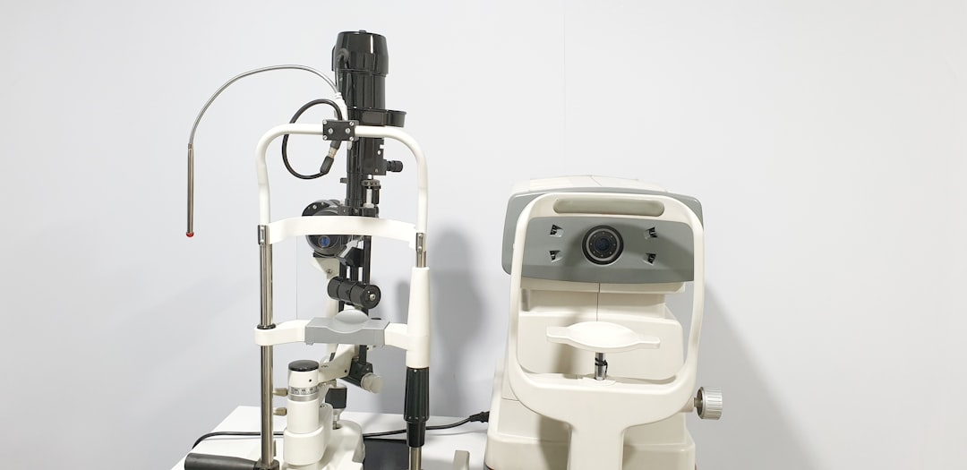
- Slit-Lamp Biomicroscopy: Using a high-magnification biomicroscope with a narrow, focused slit beam of light to view optical cross-sections of the cornea, anterior chamber, and crystalline lens. This allows your optometrist to grade the density, type, and anatomical depth of any opacities.
- Retinal Health Evaluation: Inspecting the macular area, optic nerve head, and peripheral blood vessels behind the clouded lens to ensure that other conditions, such as macular degeneration or glaucoma, are not contributing to vision changes.
Understanding what takes place during an evaluation helps demystify the process; you can read our detailed breakdown in comprehensive eye test 101. Please remember to bring your current spectacles and your Medicare card to your consultation.
If you ever experience sudden visual disturbances, such as abrupt blurring, flashes of light, or ocular discomfort, arrange prompt professional assessment. You can easily Book an Eye Test or reach out via our Contact Us page to have your ocular health thoroughly assessed.
Practical Solutions for Managing Cataracts and Supporting Your Daily Sight
Receiving a cataract diagnosis does not mean you require immediate surgery. Cataracts develop slowly, and management is guided by how significantly the lens changes impact your daily comfort, hobbies, and independence.
In the early and moderate stages, conservative, non-surgical approaches can effectively support clear vision:
- Updated Spectacle Scripts: Fine-tuning your spectacles/glasses with specialised anti-reflective coatings can improve contrast and minimise surface glare.
- Tailored Task Lighting: Utilising direct, positionable lamps with clean, white light for reading, cooking, and close hobbies reduces eye fatigue.
- Specialised Optical Aids: Incorporating handheld magnifiers or specialised lenses (including prism where relevant) can assist with detailed close work.
- Outdoor Glare Reduction: Wearing polarised or tinted lenses outdoors helps manage light sensitivity and improves comfort in bright conditions.
For additional insights on maintaining long-term ocular wellbeing, explore a practical guide to long-term eye health.
Specialist Referral and Modern Surgical Management
When lens clouding progresses to a stage where non-surgical solutions no longer provide adequate visual clarity for driving, reading, or everyday tasks, your optometrist will arrange a referral to an ophthalmologist for surgical consideration.
Cataract surgery is a standard, highly refined day procedure. According to the HealthyWA cataract surgery guidelines, the operation is commonly performed under local anaesthesia.
The standard surgical technique is phacoemulsification:
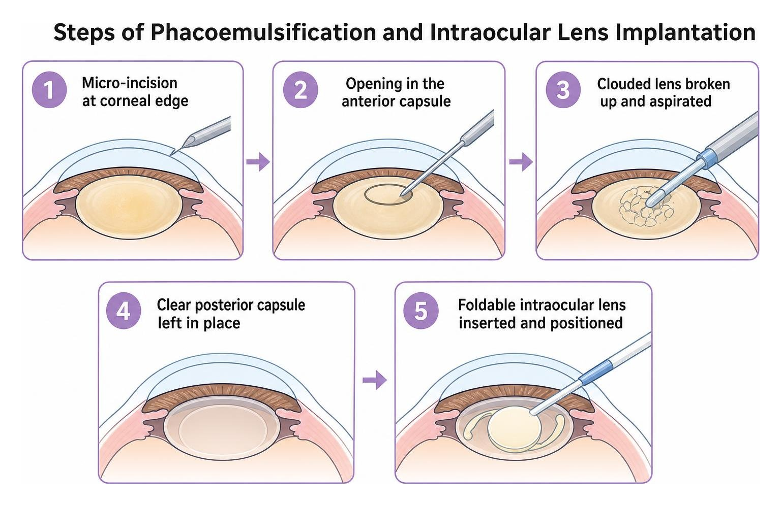
- The ophthalmologist creates a tiny micro-incision at the corneal border.
- A circular opening is made in the front membrane of the lens capsule.
- An ultrasonic instrument gently breaks the cloudy natural lens into tiny fragments, which are carefully removed.
- The clear posterior capsule remains in place to support the new artificial lens.
- A foldable intraocular lens (IOL) is inserted through the micro-incision and positioned securely within the capsular bag.
There are several intraocular lens options available depending on your visual goals:
- Monofocal IOLs: Provide sharp focus at a single set distance (usually distance vision). Spectacles/glasses are typically used for reading or computer work. Monovision lenses are an available option where one eye is set for distance and the other for near vision.
- Toric IOLs: Feature built-in cylindrical correction to correct corneal astigmatism alongside cataract removal.
- Extended Depth of Focus (EDOF) and Multifocal IOLs: Advanced optical designs that distribute light across multiple focal points, potentially reducing the need for reading spectacles.
Optometrists play a central role in collaborative co-management alongside ophthalmic surgeons. We conduct comprehensive pre-operative assessments and provide attentive post-operative reviews to check intraocular pressure, monitor tissue healing, and update your final spectacle script. Learn more about how we support patients through eye conditions – cataracts.
Can You Slow Down Cataract Development and Protect Your Eyes?
While age-related lens changes cannot be entirely eliminated, evidence-based lifestyle habits may help slow the progression of lens opacification and protect your overall ocular health:
- Broad-Spectrum UV Protection: Wear sunglasses that meet the Australian Standard for UV protection, ensuring they provide maximum defence against UVA and UVB radiation. Pairing sunglasses with a wide-brimmed hat provides excellent defence against high ambient UV levels.
- Smoking Cessation: Quitting smoking eliminates a major source of systemic free radicals, reducing oxidative damage to crystallin proteins.
- Nutritional and Dietary Support: Consume a balanced, nutrient-dense diet rich in antioxidants, lutein, zeaxanthin, vitamin C, vitamin E, and omega-3 fatty acids. Leafy green vegetables, colourful berries, nuts, and fish support cellular health across ocular tissues. For further reading, see a practical guide to long-term eye health.
- Glycaemic Management: If you have diabetes, maintaining stable blood glucose levels under the guidance of your General Practitioner (GP) helps protect the crystalline lens from osmotic stress and sugar-induced clouding.
- Eye Protection: Use certified safety eyewear during sports, gardening, and home maintenance to prevent mechanical eye trauma.
Regular visual reviews allow your optometrist to monitor subtle lens changes over time. Adults over 65 should have an annual evaluation, while younger adults are advised to attend every two years. Review our range of services under eye tests to stay proactive about your ocular wellbeing.
Empowering Your Visual Journey with Attentive Optometric Guidance
Understanding your ocular health allows you to navigate changes in your sight with confidence and peace of mind. By recognising the early signs of crystalline lens changes, safeguarding your eyes against UV exposure, and attending regular visual evaluations, you can actively support your visual clarity for years to come.
Whether you have noticed new glare when driving at night, feel your current spectacles/glasses are no longer performing well, or simply wish to have your lens clarity comprehensively reviewed, our team is dedicated to providing unhurried, reassuring care where you are truly listened to and supported at every stage.
To have your eyes thoroughly evaluated, please Book an Eye Test or reach out through our Contact Us page today. Please remember to bring your current spectacles/glasses and your Medicare card to your appointment.
Frequently Asked Questions About Cataracts
Can a cataract spread from one eye to the other?
No. Cataracts cannot spread from one eye to the other. A cataract is an isolated internal breakdown of proteins within the crystalline lens of a specific eye, rather than an infection or transmissible condition. However, because both eyes experience similar biological ageing, UV exposure, and systemic health influences, cataracts frequently develop in both eyes around the same stage of life, though the rate of progression may differ between each eye.
Can eye drops, exercises, or diet dissolve an existing cataract?
No. There are currently no scientifically proven eye drops, medications, or visual exercises that can dissolve or reverse an existing cataract once lens proteins have denatured and aggregated. While nutritious dietary habits and UV protection can help support general lens health and may slow further progression, the only definitive method to remove a developed cataract is through surgical extraction by an ophthalmologist.
What is an ‘after-cataract’ and how is it managed?
An ‘after-cataract’ is the informal term for Posterior Capsule Opacification (PCO). During modern cataract surgery, the natural posterior capsule is preserved to support the artificial intraocular lens. In some individuals, residual microscopic lens cells can migrate across this capsule months or years later, causing the membrane to become hazy and recreating misty symptoms.
PCO is easily managed by an ophthalmologist using a quick, painless outpatient procedure known as a YAG laser capsulotomy. The laser creates a small central opening in the cloudy capsule, restoring a clear path for light to reach the retina without requiring surgical incisions.